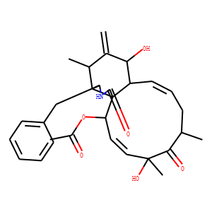
| IUPAC Name | [(1R,2R,3E,5R,7S,9E,11R,12S,14S,15R,16S)-16-benzyl-5,12-dihydroxy-5,7,14-trimethyl-13-methylidene-6,18-dioxo-17-azatricyclo[9.7.0.01,15]octadeca-3,9-dien-2-yl] acetate |
| InChI | InChI=1S/C30H37NO6/c1-17-10-9-13-22-26(33)19(3)18(2)25-23(16-21-11-7-6-8-12-21)31-28(35)30(22,25)24(37-20(4)32)14-15-29(5,36)27(17)34/h6-9,11-15,17-18,22-26,33,36H,3,10,16H2,1-2,4-5H3,(H,31,35)/b13-9+,15-14+/t17-,18+,22-,23-,24+,25-,26+,29+,30+/m0/s1 |
| Reference | 1. Curr Mol Med. 2019;20(1):79-88. doi: 10.2174/1566524019666191007104816.
<br>
Cytochalasin D Promotes Osteogenic Differentiation of MC3T3-E1 Cells via
p38-MAPK Signaling Pathway.
<br>
Liu Q(1), Zhuang Y(1), Ouyang N(2), Yu H(1).
<br>
Author information:<br>
(1)Department of Oral and Cranio-maxillofacial Surgery, Shanghai Ninth People’s
Hospital, College of Stomatology, Shanghai Jiao Tong University School of
Medicine; Shanghai Key Laboratory of Stomatology & Shanghai Research Institute
of Stomatology, National Clinical Research Center for Oral Diseases, Shanghai
200011, China.<br>
(2)Department of Orthodontics, Shanghai Ninth People’s Hospital, College of
Stomatology, Shanghai Jiao Tong University, School of Medicine, Shanghai 200011,
China.
<br>
BACKGROUND: Bone defect caused by trauma, tumor resection, infection or
congenital malformation is a common clinical disease. Bone tissue engineering is
regarded as a promising way of bone defect reconstruction. Thus, agents that can
promote osteogenesis have received great attention. Cytochalasin D (Cyto D), a
metabolite derived from molds, proves to be able to modify actin, reorganize
cytoskeleton, and then promote the osteogenic differentiation.
OBJECTIVE: The purpose of this study was to explore the effect and mechanism of
Cyto D on osteogenic differentiation of mouse pre-osteoblast MC3T3-E1 cells.
METHODS: The optimum concentration of Cyto D was explored. The osteogenic
differentiation of MC3T3-E1 cells induced by Cyto D was assessed by alkaline
phosphatase (ALP) staining, Alizarin Red S (ARS) staining, western blotting and
quantitative real-time polymerase chain reaction (RT-qPCR). In addition, a
specific pathway inhibitor was utilized to explore whether MAPK pathways were
involved in this process.<br>
RESULTS: The results showed that the optimized concentration of action was
10-2µg/ml. The expression of Runx2, OCN and OSX was up-regulated by the
supplement of Cyto D. ALP activity, calcium deposition, and phosphorylation
level of p38 protein were also improved. Inhibition of the pathway significantly
reduced the activation of p38, and the expression of osteogenic-related genes.
CONCLUSION: Cyto D can promote the osteogenic differentiation of MC3T3 cells via
the p38-MAPK signaling pathway, but not the ERK1/2 or JNK, and it is a potential
agent to improve the osteogenesis of MC3T3 cells.
<br><br>
2. J Cell Biol. 1982 Jan;92(1):79-91. doi: 10.1083/jcb.92.1.79.
<br>
Action of cytochalasin D on cytoskeletal networks.
<br>
Schliwa M.
<br>
Extraction of SC-1 cells (African green monkey kidney) with the detergent Triton
X-100 in combination with stereo high-voltage electron microscopy of whole mount
preparations has been used as an approach to determine the mode of action of
cytochalasin D on cells. The cytoskeleton of extracted BSC-1 cells consists of
substrate-associated filament bundles (stress fibers) and a highly cross-linked
network of four major filament types extending throughout the cell body; 10-nm
filaments, actin microfilaments, microtubules, and 2- to 3-nm filaments. Actin
filaments and 2- to 3-nm filaments form numerous end-to-side contacts with other
cytoskeletal filaments. Cytochalasin D treatment severely disrupts network
organization, increases the number of actin filament ends, and leads to the
formation of filamentous aggregates or foci composed mainly of actin filaments.
Metabolic inhibitors prevent filament redistribution, foci formation, and cell
arborization, but not disorganization of the three-dimensional filament network.
In cells first extracted and then treated with cytochalasin D, network
organization is disrupted, and the number of free filament ends is increased.
Supernates of preparations treated in this way contain both short actin
filaments and network fragments (i.e., actin filaments in end-to-side contact
with other actin filaments). It is proposed that the dramatic effects of
cytochalasin D on cells result from both a direct interaction of the drug with
the actin filament component of cytoskeletal networks and a secondary cellular
response. The former leads to an immediate disruption of the ordered
cytoskeletal network that appears to involve breaking of actin filaments, rather
than inhibition of actin filament-filament interactions (i.e., disruption of
end-to-side contacts). The latter engages network fragments in an
energy-dependent (contractile) event that leads to the formation of filament
foci.
<br><br>
3. Asian Pac J Trop Med. 2012 Mar;5(3):169-74. doi: 10.1016/S1995-7645(12)60019-4.
<br>
Cytochalasin D, a tropical fungal metabolite, inhibits CT26 tumor growth and
angiogenesis.
<br>
Huang FY(1), Li YN, Mei WL, Dai HF, Zhou P, Tan GH.
<br>
Author information:<br>
(1)Agriculture College, Hainan University, Haikou, China.
<br>
OBJECTIVE: To investigate whether cytochalasin D can induce antitumor activities
in a tumor model.<br>
METHODS: Murine CT26 colorectal carcinoma cells were cultured in vitro and
cytochalasin D was used as a cytotoxic agent to detect its capabilities of
inhibiting CT26 cell proliferation and inducing cell apoptosis by MTT and a
TUNEL-based apoptosis assay. Murine CT26 tumor model was established to observe
the tumor growth and survival time. Tumor tissues were used to detect the
microvessel density by immunohistochemistry. In addition, alginate encapsulated
tumor cell assay was used to quantify the tumor angiogenesis in vivo.
RESULTS: Cytochalasin D inhibited CT26 tumor cell proliferation in time and dose
dependent manner and induced significant CT26 cell apoptosis, which almost
reached the level induced by the positive control nuclease. The optimum
effective dose of cytochalasin D for in vivo therapy was about 50 mg/kg.
Cytochalasin D in vivo treatment significantly inhibited tumor growth and
prolonged the survival times in CT26 tumor-bearing mice. The results of
immunohistochemistry analysis and alginate encapsulation assay indicated that
the cytochalasin D could effectively inhibited tumor angiogenesis.
CONCLUSIONS: Cytochalasin D inhibits CT26 tumor growth potentially through
inhibition of cell proliferation, induction of cell apoptosis and suppression of
tumor angiogenesis.
<br>
|