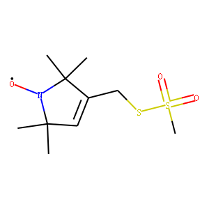
| Reference | [1]. Appl Magn Reson. 2018 Dec;49(12):1385-1395. doi: 10.1007/s00723-018-1070-6. Epub 2018 Nov 14.<br />
Nucleotide Spin Labeling for ESR Spectroscopy of ATP-Binding Proteins.<br />
Muok AR(1), Chua TK(1), Le H(1), Crane BR(1).<br />
Author information: (1)Department of Chemistry and Chemical Biology, Cornell University Ithaca, NY.<br />
Site-directed spin labeling of proteins by chemical modification of engineered cysteine residues with the molecule MTSSL (1-Oxyl-2,2,5,5-tetramethylpyrroline-3-methyl methanethiosulfonate) has been an invaluable tool for conducting double electron electron resonance (DEER) spectroscopy experiments. However, this method is generally limited to recombinant proteins with a limited number of reactive Cys residues that when modified will not impair protein function. Here we present a method that allows for spin-labeling of protein nucleotide binding sites by adenosine diphosphate (ADP) modified with a nitroxide moiety on the β-phosphate (ADP-β-S-SL). The synthesis of this ADP analog is straightforward and isolation of pure product is readily achieved on a standard reverse-phase high-performance liquid chromatography (HPLC) system. Furthermore, analyses of isolated ADP-β-S-SL by LC-mass spectrometry confirm that the molecule is extremely stable under ambient conditions. The crystal structure of ADP-β-S-SL bound to the ATP pocket of the histidine kinase CheA reveals specific targeting of the probe, whose nitroxide moiety is mobile on the protein surface. Continuous wave and pulsed ESR measurements demonstrate the capability of ADP-β-S-SL to report on active site environment and provide reliable DEER distance constraints.<br />
DOI: 10.1007/s00723-018-1070-6 PMCID: PMC6342010 PMID: 30686862<br />
<br />
[2]. Biophys J. 2001 Jun;80(6):2867-85. doi: 10.1016/S0006-3495(01)76253-4.<br />
High-resolution probing of local conformational changes in proteins by the use of multiple labeling: unfolding and self-assembly of human carbonic anhydrase II monitored by spin, fluorescent, and chemical reactivity probes.<br />
Hammarström P(1), Owenius R, Mårtensson LG, Carlsson U, Lindgren M.<br />
Author information: (1)Department of Chemistry, Linköping University, SE-581 83 Linköping, Sweden.<br />
Two different spin labels, N-(1-oxyl-2,2,5,5-tetramethyl-3-pyrrolidinyl)iodoacetamide (IPSL) and (1-oxyl-2,2,5,5-tetramethylpyrroline-3-methyl) methanethiosulfonate (MTSSL), and two different fluorescent labels 5-((((2-iodoacetyl)amino)-ethyl)amino)naphtalene-1-sulfonic acid (IAEDANS) and 6-bromoacetyl-2-dimetylaminonaphtalene (BADAN), were attached to the introduced C79 in human carbonic anhydrase (HCA II) to probe local structural changes upon unfolding and aggregation. HCA II unfolds in a multi-step manner with an intermediate state populated between the native and unfolded states. The spin label IPSL and the fluorescent label IAEDANS reported on a substantial change in mobility and polarity at both unfolding transitions at a distance of 7.4-11.2 A from the backbone of position 79. The shorter and less flexible labels BADAN and MTSSL revealed less pronounced spectroscopic changes in the native-to-intermediate transition, 6.6-9.0 A from the backbone. At intermediate guanidine (Gu)-HCl concentrations the occurrence of soluble but irreversibly aggregated oligomeric protein was identified from refolding experiments. At approximately 1 M Gu-HCl the aggregation was found to be essentially complete. The size and structure of the aggregates could be varied by changing the protein concentration. EPR measurements and line-shape simulations together with fluorescence lifetime and anisotropy measurements provided a picture of the self-assembled protein as a disordered protein structure with a representation of both compact as well as dynamic and polar environments at the site of the molecular labels. This suggests that a partially folded intermediate of HCA II self-assembles by both local unfolding and intermolecular docking of the intermediates vicinal to position 79. The aggregates were determined to be 40-90 A in diameter depending on the experimental conditions and spectroscopic technique used.<br />
DOI: 10.1016/S0006-3495(01)76253-4 PMCID: PMC1301471 PMID: 11371460<br />
<br />
[3]. Structure. 2009 Sep 9;17(9):1187-94. doi: 10.1016/j.str.2009.07.011.<br />
The orientation of a tandem POTRA domain pair, of the beta-barrel assembly protein BamA, determined by PELDOR spectroscopy.<br />
Ward R(1), Zoltner M, Beer L, El Mkami H, Henderson IR, Palmer T, Norman DG.<br />
Author information: (1)College of Life Sciences, University of Dundee, Dundee, UK.<br />
The outer membrane beta-barrel trans-membrane proteins in gram-negative bacteria are folded into the membrane with the aid of polypeptide transport-associated (POTRA) domains. These domains occur, and probably function, as a tandem array situated on the periplasmic side of the outer membrane. Two crystal structures and one NMR study have attempted to define the structure and articulation of the POTRA domains of the Escherichia coli, prototypic Omp85 protein BamA. We have used pulsed electron paramagnetic resonance (EPR) to determine the distance and distance distribution between (1-Oxyl-2,2,5,5-tetramethylpyrroline-3-methyl) methanethiosulfonate spin labels (MTSSL), placed across the domain interface of the first two POTRA domains of BamA. Our results show tightly defined interdomain distance distributions that indicate a well-defined domain orientation. Examination of the known structures revealed that none of them fitted the EPR data. A combination of EPR and NMR data was used to generate converged structures with defined domain-domain orientation.<br />
DOI: 10.1016/j.str.2009.07.011 PMID: 19748339
|

